
Exercise 12 microscopic anatomy and organization of skeletal muscle – Exercise 12: Microscopic Anatomy and Organization of Skeletal Muscle embarks on an in-depth exploration of the intricate structure and remarkable function of skeletal muscle. This journey unveils the fundamental components that orchestrate movement, revealing the interplay of fascicles, fibers, myofibrils, and myofilaments.
Prepare to delve into the intricacies of muscle architecture, unlocking the secrets of muscle contraction and neuromuscular communication.
As we delve deeper into this topic, we will dissect the sarcomere, the fundamental unit of muscle contraction, unraveling the roles of the Z-line, M-line, and H-zone. The sliding filament theory will take center stage, illuminating the dynamic dance of actin and myosin filaments that generates force.
Microscopic Anatomy of Skeletal Muscle
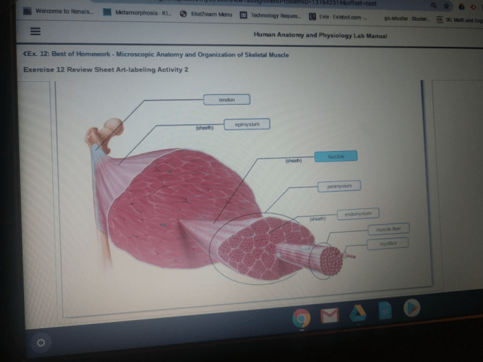
Skeletal muscle is a highly organized tissue composed of several hierarchical levels. The largest unit is the muscle itself, which is composed of bundles of fascicles. Each fascicle is surrounded by a connective tissue sheath called the perimysium and contains numerous muscle fibers.
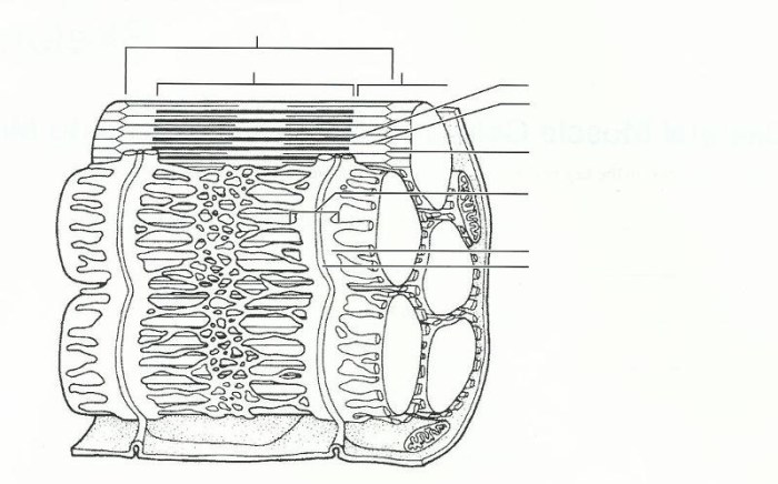
Muscle fibers are long, cylindrical cells that contain myofibrils, which are composed of repeating units called sarcomeres. Sarcomeres are the basic contractile units of muscle and contain the protein filaments actin and myosin.
Sarcolemma, Endomysium, Perimysium, and Epimysium
The sarcolemma is the plasma membrane of the muscle fiber and is responsible for maintaining the cell’s integrity and excitability. The endomysium is a thin layer of connective tissue that surrounds each muscle fiber and provides support. The perimysium is a thicker layer of connective tissue that surrounds each fascicle and provides additional support.
The epimysium is the outermost layer of connective tissue that surrounds the entire muscle and provides protection.
Sarcomere Structure and Function: Exercise 12 Microscopic Anatomy And Organization Of Skeletal Muscle
The sarcomere is the basic contractile unit of skeletal muscle. It is composed of two types of protein filaments: actin and myosin. Actin filaments are thin and contain a myosin-binding site, while myosin filaments are thick and contain a head region that can bind to actin.
The sarcomere is organized into repeating units called A-bands, I-bands, H-zones, and Z-lines.
Role of the Z-line, M-line, and H-zone in Muscle Contraction, Exercise 12 microscopic anatomy and organization of skeletal muscle
The Z-line is a dense protein structure that anchors the actin filaments at the ends of the sarcomere. The M-line is a less dense protein structure that anchors the myosin filaments in the middle of the sarcomere. The H-zone is the region of the sarcomere that contains only myosin filaments.
Sliding Filament Theory of Muscle Contraction
The sliding filament theory of muscle contraction states that muscle contraction occurs when the actin and myosin filaments slide past each other. This sliding is caused by the interaction of the myosin heads with the actin-binding sites. As the myosin heads bind to the actin-binding sites, they undergo a conformational change that causes them to pull the actin filaments towards the center of the sarcomere.
This sliding of the filaments causes the sarcomere to shorten and the muscle to contract.
Neuromuscular Junction
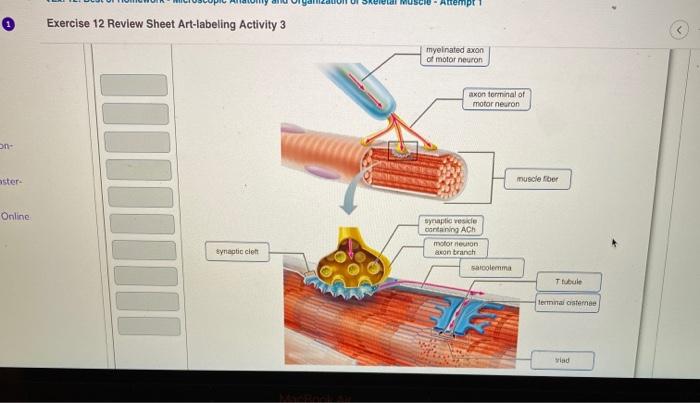
The neuromuscular junction is the site of communication between a motor neuron and a muscle fiber. The motor neuron releases acetylcholine, which binds to receptors on the surface of the muscle fiber. This binding causes the muscle fiber to depolarize, which triggers the release of calcium ions from the sarcoplasmic reticulum.
The calcium ions bind to troponin, which causes a conformational change that exposes the myosin-binding sites on the actin filaments. This allows the myosin heads to bind to the actin-binding sites and initiate muscle contraction.
Muscle Fiber Types
There are three main types of muscle fibers: slow-twitch, fast-twitch, and intermediate. Slow-twitch fibers are fatigue-resistant and are used for sustained activities, such as walking and running. Fast-twitch fibers are fatigue-prone and are used for short, powerful activities, such as sprinting and jumping.
Intermediate fibers have characteristics of both slow-twitch and fast-twitch fibers.
Characteristics, Functions, and Metabolic Properties of Each Fiber Type
Slow-twitch fibers are characterized by a low myosin ATPase activity, a high mitochondrial content, and a high oxidative capacity. Fast-twitch fibers are characterized by a high myosin ATPase activity, a low mitochondrial content, and a low oxidative capacity. Intermediate fibers have characteristics that are intermediate between slow-twitch and fast-twitch fibers.
Examples of How Different Muscle Fiber Types Are Adapted to Specific Activities
Slow-twitch fibers are adapted to sustained activities because they are fatigue-resistant and have a high oxidative capacity. Fast-twitch fibers are adapted to short, powerful activities because they have a high myosin ATPase activity and a low oxidative capacity. Intermediate fibers are adapted to activities that require both sustained and powerful contractions.
Muscle Adaptations to Exercise
Exercise can influence muscle fiber size, number, and type. Hypertrophy is an increase in muscle fiber size, which occurs in response to resistance training. Hyperplasia is an increase in muscle fiber number, which occurs in response to endurance training. Fiber type conversion is a change in muscle fiber type, which occurs in response to changes in training intensity and duration.
Role of Hypertrophy, Hyperplasia, and Fiber Type Conversion in Muscle Adaptation
Hypertrophy increases muscle strength and power, while hyperplasia increases muscle endurance. Fiber type conversion can improve muscle performance by increasing the proportion of fast-twitch fibers, which are more powerful than slow-twitch fibers.
Common Queries
What is the basic unit of muscle contraction?
The sarcomere is the fundamental unit of muscle contraction.
What is the role of the Z-line in muscle contraction?
The Z-line anchors the ends of actin filaments and marks the boundaries of the sarcomere.
How does the sliding filament theory explain muscle contraction?
The sliding filament theory proposes that actin and myosin filaments slide past each other during muscle contraction, causing the sarcomere to shorten.
What are the different types of muscle fibers?
There are three main types of muscle fibers: slow-twitch, fast-twitch, and intermediate.
How does exercise affect muscle fiber size and type?
Exercise can influence muscle fiber size, number, and type through mechanisms such as hypertrophy, hyperplasia, and fiber type conversion.
